Department of Integrated Neuro-Oncology and RadioneurosurgeryCall +380 44 483-95-73, +380 44 483-94-13, +380 44 483-70-99, +380 44 483-70-89 E-mail: cho72@ukr.net How to find: Institute of Neurosurgery, Building 7, 2nd floor (see Map and Institute scheme). 
Olga Y. Chuvashova
Head of the department, MD, PhD, DSc, Professor, 
Oleksandr Ya. Hlavatskyi
Leading Expert in Neurosurgery, 
Oksana V. Zemskova
Chief Researcher, 
Dmytro S. Teslenko
Researcher, 
Iryna V. Kruchok
Researcher, 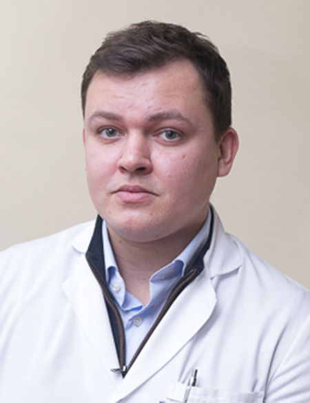
Valerii V. Cheburakhin
Researcher, 
Yuliia V. Medvedovska
Researcher 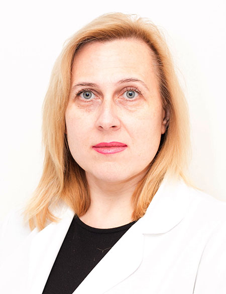
Iryna M. Shuba
Junior Researcher, The history of the newly established Department of Integrated Neuro-Oncology and Radioneurosurgery is associated with the institutional restructuring implemented on 1 June 2026, as well as with the rapid advancement of neuroradiology, stereotactic radiosurgery, and multidisciplinary approaches to the management of central nervous system (CNS) disorders, particularly neuro-oncological diseases. Given the inseparable diagnostic and therapeutic continuum in modern neuro-oncology and neurosurgery—from advanced neuroimaging and treatment planning to radiosurgical, systemic, and combined therapies—it became both appropriate and necessary to integrate the neuroradiological, radioneurosurgical, and adjuvant treatment services into a single multidisciplinary department. This organizational model is consistent with the internationally recognized concept of a Comprehensive Neuro-Oncology & Radiosurgery Center, as promoted by the European Association of Neuro-Oncology (EANO), the Society for Neuro-Oncology (SNO), the International Stereotactic Radiosurgery Society (ISRS), and the European Society for Radiotherapy and Oncology (ESTRO). It ensures continuity of specialized care for patients with CNS tumors, vascular and functional disorders, optimizes the utilization of high-technology medical equipment, and integrates clinical protocols, scientific research, and educational activities within a unified organizational framework. The history of the Department began in April 2005 with the establishment of the Department of Neuroimaging Studies, which included an MRI unit. Between 2008 and 2010, a new building was constructed specifically to house a linear accelerator and a complex of equipment necessary for radio-neurosurgery. On June 16, 2010, the Department of Neuroradiology and Radioneurosurgery was officially opened. In 2010, the Department of Neuroimaging Studies was merged with the newly created Department of Neuroradiology and Radioneurosurgery, forming a single unit — the Department of Neuroradiology and Radioneurosurgery. The Department is equipped with: – a 1.5 Tesla MRI scanner “Philips Intera 1.5T”; – a multislice CT scanner “Brilliance CT 64-slice” (Philips, Netherlands); – a linear accelerator “Trilogy” (Varian, USA) with a stereotactic system “BrainLab.” The main areas of scientific and practical work of the department
– development of integrated neuroimaging methods to improve the effectiveness of diagnosis and surgical treatment of space-occupying lesions and brain tumors (metastases, vestibular schwannomas [neurinomas], meningiomas, paragangliomas, trigeminal schwannomas, astrocytomas, glioblastomas, pituitary adenomas, craniopharyngiomas, ependymomas, lymphomas, hemangioblastomas, arteriovenous malformations, cavernous angiomas); – implementation of stereotactic radiosurgical treatment of primary and secondary tumor and vascular brain lesions (metastases, vestibular schwannomas [neurinomas], meningiomas, paragangliomas, trigeminal schwannomas, astrocytomas, glioblastomas, pituitary adenomas, craniopharyngiomas, ependymomas, lymphomas, hemangioblastomas, arteriovenous malformations, cavernous angiomas); – application of stereotactic radiotherapy in the treatment of CNS oncological pathologies with an individualized patient-centered approach; – conducting functional MRI studies in various types of focal brain lesions; – ensuring MRI guidance in the planning of stereotactic biopsy and developing criteria for target selection and trajectory planning; – using MSCT-perfusion studies for monitoring oncology patients and patients after radioneurosurgical interventions; – performing diffusion MRI studies in vascular and oncological brain pathologies; – carrying out MR tractography for visualization of major brain pathways; – development of a set of techniques and specific technological approaches for highly effective computer planning of local interventions, followed by extrapolation and integration of the obtained data into navigational operating systems; – improvement of neuroimaging studies in spinal pathology, evaluation of outcomes of modern surgical treatments of spinal disorders, as well as research into vascular and inflammatory CNS processes and orphan diseases. Updated 05 July 2026 |

 orcid.org/0000-0001-6767-4665