Neuroradiology DepartmentTel. +380 66 673 00 96 Liudmyla Y. Boyko E-mail: robakoleg@ukr.net How to find: Institute of Neurosurgery, Building 6, 2nd floor (see Map and Institute scheme). 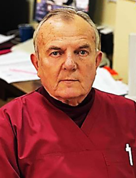
Oleg P. Robak
Head of department MD, Radiology 
Stanislav V. Makhovsky
MD, Radiology 
Olga Y. Harmatina
MD, Radiology 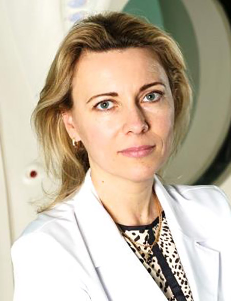
Ivanna L. Yakovenko
MD, Radiology 
Darya O. Nebylitsa
MD, Radiology 
Iryna V. Sydorak
MD, Radiology 
Olesya Y. Pylypas
MD, Radiology 
Andriy V. Kravchuk
MD, Radiology The department of neuroradiology specializes in the diagnosis of craniocerebral trauma, neurooncology, vascular pathology of the brain and spinal cord. Annually, the department performs about 15,000 studies, including about 11,500 – using special methods.
It is used in the department: Open low-field magnetic resonance imaging 0.5 T(about 2,000 studies per year), which is especially important for patients with a fear of closed spaces: Native MRI of the brain MRI of the brain with intravenous enhancement MRI with focused visualization of cranial nerves MRI with focused visualization of orbital structures MRI with focused visualization of the structures of the sellar area MRI with DWI MRI with the tirm program MRI of the cervical spine MRI of the cervical spinal cord MR myelography of the cervical region MRI of the neck with intravenous enhancement MRI of the thoracic spine MRI of the thoracic spinal cord MR-myelography of the thoracic department MRI of the chest with intravenous enhancement MRI of the lumbosacral spine MRI of the spinal cord of the lumbar spine MR myelography of the lumbar region MRI of the lumbar region with intravenous enhancement Multi-detector computed tomography on 160-slice CT (about 8 thousand studies per year) Native MDCT of the head MDCT of the head with intravenous enhancement (2 studies) MDCT of the head with enhancement through an injector (2 studies) Cerebral MDCT-AG (2 studies) Cerebral MDCT-AG with delayed venous phase (3 studies) Cerebral MDCT perfusion (2 studies) MDCT cisternography MDKT navigation MDCT of the temporal bone MDCT of the facial skull MDCT of the paranasal sinuses MDCT-AG of the neck (2 studies) MDCT-AG of the head and neck (2 studies) MDCT of the cervical spine MDCT-AG of extracranial vessels (2 studies) MDCT of the thoracic spine MDCT of the lumbar spine MDCT of pelvic bones MDCT of the chest cavity MDCT of the joints MDCT of the extremities MDCT oncoscreening (4 studies) Digital radiography: Skull in 2 projections Nasal bones in 2 projections Sacrificial sinuses in 2 projections Cervical spine in 2 projections Cervical spine with functional samples (3 projections) X-ray of the Turkish saddle Thoracic spine in 2-projections Thoracic section of the spine with functional samples (3 projections) Lumbar section of the spine in 2 projections Lumbar section of the spine with functional samples (3 projections) Coccyx in 2 projections Bones of the pelvis and hip joints in 2 projections Collarbones in 2 projections Shoulder blade in 2 projections Bones of the upper limbs in 2 projections Bones of the lower limbs in 2 projections Knee joints in 2 projections Shoulder joints in 2 projections Elbow joints in 2 projections Ankle joints in 2 projections Chest cavity organs in 2 projections Abdominal organs in 2 projections Digital angiography of the brain and spinal cord on the most modern angiograph (about 2,000 studies per year) Updated 26 August 2025 |